Contacts
Motivation
Telesonography is a term that describes the application of telemedicine in Ultrasound (US) field. Although US imaging has a unique combination of advantages, like its safety in use, non-invasiveness, precautions-free scanning routine, and ability to image soft tissues and detecting flow, it is until today limited in use to hospitals due to the high dependancy on a trained sonographer to perform the scan. 3D US removes this dependancy by relaxing the probe positioning constrains giving the ability to anyone to operate the scan. However, today’s 3D systems suit only well-equipped hospitals due to their stationary size, high cost, and considerable power consumption, which are the result of the tremendous computation requirements of volumetric reconstruction. Therefore, telesonography is until today just a dream, and US is not served in many situations in need, like remote isolated areas, special environments (space stations, aircrafts, battlefields, etc.), rescue situations, and underdeveloped regions.
Goal
In order to solve this vicious cycle and allow the usage of US by anyone anywhere, a telesonography-capable system should be designed featuring: 3D imaging, portability, battery-range power consumption, good quality imaging, and desirably, low in cost. This has formulated our objective; a telesonography-capable back-end digital processing system.
Achievements
A considerable step towards unlocking telesonography has been successfully achieved by designing an unprecedented single-FPGA 3D medical US imaging system with a 5 W consumption. Our imaging system features: (i) 1024 channel of independent information processing (state-of-the-art), (ii) two input means: realtime data via optical connection with a US transducer, and offline data via Ethernet, (iii) complete digital processing platform from the receive of the digitized raw data until the rendering of the reconstruction on a screen, (iv) three common US imaging modes for image enhancement flexibility at the cost of the frame rate, (v) extreme scalability: downscaling for 2D imaging, or further upscaling on a resources-capable FPGA, and (vi) high-definition live video output of the scans. We have developed a prototype whose size is currently 26.7cm × 14cm × 0.16cm. The material cost is less than 4000$, with a path to further reduction by implementing a custom board around the FPGA for mass production, instead of the off-the- shelf development board. This compares very favorably to current commercial 3D systems, which cost ∼100K$ for the imaging system only.
This has been achieved through three main phases:
- Algorithmic innovation, development, and investigation on the Matlab framework
- Hardware realization of carefully selected algorithms on an FPGA-based embodiment by Xilinx Inc.
- Extensive and intensive assessment for the platform in terms of the resource utilization, reconstruction quality, portability, cost, and even system flexibility and scalability, on both Matlab and Xilinx Vivado
Our contributions have been consolidated to successfully achieve the aimed target of a first telesonography-capable prototype allowing the usage of medical US anywhere and by any personnel, with major societal benefits.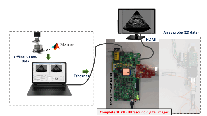
Acknowledgement
This work is part of UltrasoundToGo project of the Nano-Tera.ch initiative funded by the Swiss Confederation.
Main Publications
- A. Ibrahim, P. A. Hager, A. Bartolini, F. Angiolini, M. Arditi, J.-P. Thiran, L. Benini, and G. De Micheli, “Towards Ultrasound Everywhere: A Portable 3D Digital Back-End Capable of Zone and Compound Imaging”, In IEEE Transactions on Biomedical Circuits and Systems (TBioCAS), vol. 12, no. 5, pp. 968-981, Oct. 2018.
- A. Ibrahim, P. A. Hager, A. Bartolini, F. Angiolini, M. Arditi, J.-P. Thiran, L. Benini, and G. De Micheli, “Efficient sample delay calculation for 2D and 3D ultrasound imaging”, In IEEE Transactions on Biomedical Circuits and Systems (TBioCAS), vol 11, no 4, pp. 815-831, August 2017.
- A. Ibrahim, W. Simon, D. Doy, E. Pignat, F. Angiolini, M. Arditi, J.-P. Thiran, and G. De Micheli, “Single-FPGA complete 3D and 2D medical ultrasound imager”, In The Proceedings of the 2017 Design and Architectures for Signal and Image Processing (DASIP 2017) Conference, Germany.
- A. Ibrahim, D. Doy, C. Loureiro, E. Pignat, F. Angiolini, M. Arditi, J.-P. Thiran, and G. De Micheli, “Inexpensive 1024-Channel 3D Telesonography System on FPGA”, in The Proceedings of the 2017 IEEE Biomedical Circuits and Systems (BioCAS 2017) Conference, Italy.
- F. Angiolini, A. Ibrahim, W. Simon, A. C. Yüzügüler, M. Arditi, J.-P. Thiran, and G. De Micheli, “1024-Channel 3D Ultrasound Digital Beamformer in a Single 5W FPGA”, In The Proceedings of the 2017 Design Automation and Test in Europe (DATE 2017) Conference, Switzerland.
- A. Ibrahim, F. Angiolini, M. Arditi, J.-P. Thiran, and G. De Micheli, “Apodization scheme for hardware-efficient beamformer”, In The Proceedings of the 12th Conference on PhD Research in Microelectronics and Electronics (PRIME 2016), Portugal.
- A. Ibrahim, P. A. Hager, A. Bartolini, F. Angiolini, M. Arditi, L. Benini, and G. De Micheli, “Tackling the bottleneck of delay tables in 3D ultrasound imaging”, In The Proceedings of the 2015 Design Automation and Test in Europe (DATE 2015) Conference, France, 2015.
The complete list of publications can be found here.
Open Access Code
- Complete matlab US imaging pipeline for 2D (linear and phased) and 3D (phased) imaging: https://github.com/ibrahimaya/ustogo-lsi
- Complete single-FPGA design including both the hardware side (two custom IPs and top-level block design) and the software side (MicroBlaze controller software and C#-based GUI): https://github.com/ibrahimaya/ustogo-lsi